Infectious Diseases of the Skin
Contents:
Bacterial skin infections may occur more frequently after bites and other wounds in the tropics, particularly when good hygiene cannot be maintained. Organisms responsible are commonly Staphylococcus aureus or Streptococcus pyogenes. Drug-resistant organisms are frequently acquired in the tropics. The presentations can include pyoderma or impetigo, abscess formation, erysipelas, cellulitis, lymphangitis, or ulceration. Furunculosis, or recurrent pyoderma, may be the result of colonization of the skin and nasal mucosa with S.
Boils may continue to occur weeks or months after a traveler returns and, if associated with S. In addition to pyoderma, cellulitis or erysipelas may complicate excoriated insect bites or any trauma to the skin. Cellulitis and erysipelas manifest as areas of skin erythema, edema, and warmth in the absence of an underlying suppurative focus. Unlike cellulitis, erysipelas lesions are raised, there is a clear line of demarcation at the edge of the lesion, and the lesions are more likely to be associated with fever. Cellulitis, on the other hand, is more likely to be associated with lymphangitis.
Another common bacterial skin infection, especially in children in the tropics, is impetigo due to S. Local care and a topical antibiotic such as mupirocin may be used, although a systemic antibiotic may be required.
Emerging antibiotic resistance among staphylococci and streptococci complicates antimicrobial options. Myiasis presents as a painful lesion similar to a boil. It is caused by infestation with the larval stage of the African tumbu fly Cordylobia anthropophaga or the Latin American bot fly Dermatobia hominis. At the center of the lesion is a small punctum that allows the larva to breathe. Extraction of the fly larva can be difficult, especially of the bot fly; extraction may be facilitated by first asphyxiating the larva, usually with an occlusive dressing or covering such as a bottle cap filled with petroleum jelly for several hours and then squeezing the larva out.
Tungiasis is caused by a sand flea Tunga penetrans. The female burrows into the skin, usually the foot, and produces a nodular, pale, subcutaneous lesion with a central dark spot.
The painful or pruritic lesion expands as the female produces eggs in her uterus. Loa loa filariasis occurs rarely in long-term travelers living in rural sub-Saharan Africa. The traveler may present with transient, migratory, subcutaneous, painful, or pruritic swellings produced by the adult nematode migration Calabar swelling. Rarely, the worm can be visualized crossing the conjunctiva of the eye or eyelid. Loiasis can be diagnosed by finding microfilariae in blood collected during daytime; however, since microfilaremia may be absent, filarial serologic tests may be helpful.
Gnathostomiasis is a nematode infection found primarily in Southeast Asia and less commonly in Africa and Latin America. Infection results from eating undercooked or raw freshwater fish. Infected travelers may experience transient, migratory, subcutaneous, pruritic, or painful swellings that may occur weeks or even years after exposure. The symptoms are due to migration of the worm through the body, and the central nervous system may be involved. Eosinophilia is common, and a definitive diagnosis is often difficult.
Dermatologic conditions of the ill returned traveler: Int J Infect Dis. Macular lesions are common and often nonspecific and may be due to drug reactions or viral exanthems. Superficial mycoses, such as tinea versicolor and tinea corporis, may also present as macular lesions. Tinea versicolor , due to Malassezia furfur previously Pityrosporum ovale , is characterized by asymptomatic hypopigmented or hyperpigmented oval, slightly scaly patches measuring 1—3 cm that are found on the upper chest, neck, and back. Treatment with topical or systemic azoles ketoconazole, fluconazole , terbinafine, or selenium sulfide present in some shampoos is recommended.
Tinea corporis ringworm may be caused by a number of different superficial fungi. The lesion is often a single lesion with an expanding red, raised ring, with a central area of clearing in the middle. The patient may not have noticed the tick bite. Leprosy Hansen disease frequently presents with hypopigmented or erythematous patches that are frequently hypoesthetic to pin prick and associated with peripheral nerve enlargement. This condition is almost exclusively found in immigrants from developing countries. Cutaneous larva migrans , a skin infection with the larval stage of dog or cat hookworm Ancylostoma spp.
A similar lesion that may be more urticarial and that rapidly progresses may be due to larva currens running larva due to cutaneous migration of filariform larvae of Strongyloides stercoralis. Lymphocutaneous spread of infection occurs when organisms spread along superficial cutaneous lymphatics, producing raised, linear, cord-like lesions; nodules or ulcers may also be found.
Examples include sporotrichosis, Mycobacterium marinum infection associated with exposure to water , leishmaniasis, bartonellosis cat-scratch disease , Nocardia infection, tularemia, melioidosis, and blastomycosis. Phytophotodermatitis is a noninfectious condition that results from interaction of natural psoralens, most commonly from spilled lime juice, and ultraviolet radiation from the sun.
The result is an exaggerated sunburn that gives rise to a linear, asymptomatic lesion that later develops hyperpigmentation. The hyper-pigmentation may take weeks or months to resolve. Ulcerated skin lesions may result from Staphylococcus infections or may be the direct result of an unseen spider bite. The necrotic ulcer of anthrax is often surrounded by edema and usually results from handling animal hides or products.
Rarely, a painless destructive ulcer with undermining edges may result from infection with Mycobacterium ulcerans Buruli ulcer. Of particular concern is the ulcer or less commonly, nodule caused by cutaneous leishmaniasis. The lesion is a chronic, usually painless ulcer unless superinfected, with heaped-up margins on exposed skin surfaces. Special diagnostic techniques are necessary to confirm the diagnosis.
Both topical and systemic treatments may be effective; the species of the infection often determines the treatment modality. If cutaneous leishmaniasis is suspected, clinicians can contact CDC at or parasites cdc. Soft tissue infections can occur after both freshwater and saltwater exposure, particularly if there is associated trauma. Puncture wounds due to fishhooks and fish spines, lacerations from inanimate objects during wading and swimming, and bites or stings from fish or other sea creatures may be the source of the trauma leading to waterborne infections.
Soft tissue infections associated with exposure to water or water-related animals include M. A variety of skin and soft tissue manifestations may occur in association with these infections, including cellulitis, abscess formation, ecthyma gangrenosum, and necrotizing fasciitis. Vibriosis infection may be especially severe in those with underlying liver disease and may manifest as a dramatic cellulitis with hemorrhagic bullae and sepsis.
In general, infections caused by these organisms may be more severe in those who are immunosuppressed. Acute infections related to aquatic injury should be treated with an antibiotic that provides both gram-positive and gram-negative coverage such as a fluoroquinolone or third-generation cephalosporin until a specific organism has been identified. Folliculitis typically develops 8—48 hours after exposure in contaminated water and consists of tender, pruritic papules, papulopustules, or nodules.
Skin & Soft Tissue Infections in Returned Travelers
Most patients have malaise, and some have low-grade fever. The condition is self-limited in 2—12 days; typically no antibiotic therapy is required. Wound infections after dog and cat bites are caused by a variety of microorganisms. Splenectomized patients are at particular risk of severe cellulitis and sepsis due to Capnocytophaga canimorsus after a dog bite.
Management of dog and cat bites includes consideration of rabies post-exposure prophylaxis, tetanus immunization, and antibiotic prophylaxis. Primary closure of puncture wounds and dog bites to the hand should be avoided. Antibiotic prophylaxis for dog bites is controversial, although most experts would treat splenectomized patients with amoxicillinclavulanate prophylactically. Management of monkey bites includes wound care, tetanus immunization, rabies postexposure prophylaxis, and consideration of antimicrobial prophylaxis.
When this happens it can become life-threatening.
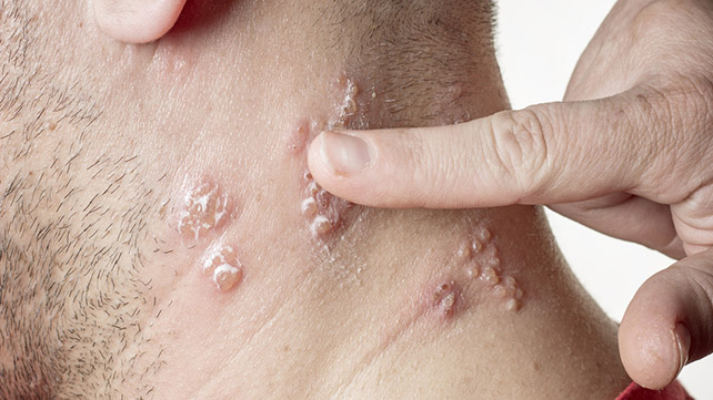
Infectious diseases of the skin.
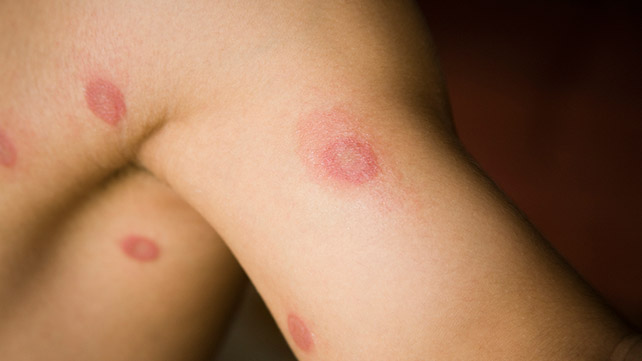
This occurs when bacteria enter the body through a break in the skin, such as a cut or a scratch. The most common viruses come from one of three groups of viruses: Body chemistry and lifestyle can increase the risk of a fungal infection.
Fungi often grow in warm, moist environments. Wearing sweaty or wet clothes is a risk factor for skin infections. A break or cut in the skin may allow bacteria to get into the deeper layers of the skin. Tiny insects or organisms burrowing underneath your skin and laying eggs can cause a parasitic skin infection. A good medical exam is the best way to determine what is causing a skin infection. Often, doctors can identify the type of skin infection based on the appearance and location.
Your doctor may ask about your symptoms and closely examine any bumps, rashes, or lesions. For example, ringworm often causes a distinct circular, scaly rash.
In other cases, a sample of skin cells can help your doctor determine the type of infection. Treatment depends on the cause of the infection and the severity. Some types of viral skin infections may improve on their own within days or weeks. Bacterial infections are often treated with topical antibiotics applied directly to the skin or with oral antibiotics. If the strain of bacteria is resistant to treatment, treating the infection may require intravenous antibiotics administered in the hospital.
You can use over-the-counter antifungal sprays and creams to treat a fungal skin infection. In addition, you can apply medicated creams to your skin to treat parasitic skin infections. Your doctor may also recommend medications to reduce discomfort like anti-inflammatory drugs. The prognosis for a skin infection varies depending on the cause. Most types of bacterial infections respond well to medications.
Contagious skin diseases
Certain strains of bacteria, such as methicillin-resistant staphylococcus aureus MRSA , are resistant to common antibiotics and are more difficult to treat. There are several ways to reduce the chances of developing a skin infection. Frequent hand washing is one of the best ways. Skin infections can vary from mild to severe. Your doctor will be able to provide the necessary treatment for recovery.
Healthline and our partners may receive a portion of revenues if you make a purchase using a link above. Scabies is a skin infestation caused by a tiny, burrowing bug. Learn about scabies symptoms, transmission, diagnosis, and treatment. Candidiasis of the skin is a fungal infection that causes a red, itchy rash.
Skin Infections
Learn about candidiasis of the skin causes, diagnosis, and treatment. We explain the basics of impetigo, a highly contagious bacterial infection of the skin. Necrotizing fasciitis is a type of soft tissue infection. It can destroy the tissue in your skin and muscles as well as subcutaneous tissue, which is….
Cellulitis is a common bacterial skin infection. Cellulitis may first appear as a red, swollen area that feels hot and tender to the touch. Athlete's foot, or tinea pedis, is a contagious fungal infection that affects the skin on the feet. This condition is contagious and can spread to the…. Infected eczema is common in people who have frequent eczema outbreaks. However, not all people with eczema will experience infections. Many people have experienced an occasional skin rash or unexplained mark.
- Be Still and Know:Incredible Hunches From Your Creator
- IWJWeekly024 (Japanese Edition)
- Piano Exercises For Dummies
- How to Pinterest (with step-by-step illustrations)
- La rivelazione dellantica carta (Leggereditore Narrativa) (Italian Edition)
- ALL YOU NEED IS LOVE: THE WAY OF JOY
- Your Kismet, my Kismet