Gamma Knife Surgery
You will return each day at your scheduled time until all fractions of the treatment plan are delivered. You will continue taking steroids and anti-seizure medications throughout treatment and for weeks thereafter. Side effects of radiation vary, depending on the tumor type, dose delivered to the tumor, number of fractions, and amount of healthy tissue in the target area.
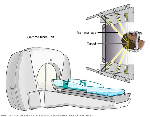
Gamma Knife radiosurgery is a very precise form of radiation therapy that focuses intense beams of gamma rays with pinpoint accuracy to treat lesions in the. Gamma Knife® is an alternative to traditional brain surgery and whole brain radiation therapy for the treatment of complex, difficult brain conditions. Leading.
Some side effects are temporary and some may be permanent. Ask your doctor about specific side effects that you may experience. General side effects may include:. Swelling edema Radiation causes tumor cells to die. Edema is extra fluid, or swelling, within the tissues of the brain. If brain swelling occurs, it can cause headaches, weakness, seizures, confusion, or speech difficulty. It may also worsen the symptoms that were present before treatment.
If you start to feel uncomfortable with headaches or any other symptoms, call your neurosurgeon or radiation oncologist. Steroid medication dexamethasone may be given to reduce brain swelling and fluid within the tumor. Steroids should always be taken with food to protect your stomach and prevent nausea. Steroids can also affect the normal bacteria in your mouth and cause a yeast infection called thrush, which appears as whitish patches on the tongue.
- Chancen und Risiken von Leveraged Buyouts für Investoren, Private-Equity-Gesellschaften und Portfoliounternehmen (German Edition).
- The Oregon Political Field Guide?
- The Which? Doctor.
- Umphrey Jacksons Recipes Especially for the Beaver Gourmet!
Do not abruptly stop taking steroids. A tapering schedule is required to avoid withdrawal. Radiation necrosis In some cases, radiosurgery may cause the center of the tumor to become necrotic dead. Radiation necrosis can happen anytime, but it most often occurs 6 to 12 months after radiosurgery.
This dying tissue can become toxic to surrounding normal tissue, and swelling may occur. Radiation necrosis may look similar to tumor regrowth on an MRI scan. Treatment for radiation necrosis may include:. After radiosurgery, MRI scans will be taken periodically so that your doctors can look for signs of response. Several months may pass before the effects of treatment are visible.
Some tumors or lesions may be completely eliminated with radiation. For others, the goal is to stop or halt the growth. For benign tumors , the goal is to stop or control the tumor's growth. For malignant primary tumors , results vary depending on the size, location, and type of tumor. Talk to your doctor about your specific prognosis. This information is not intended to replace the medical advice of your health care provider.
Stereotactic radiosurgery - Gamma Knife
Our neurosurgeons collaborate with neuroradiologists, pathologists, medical oncologists, radiation oncologists, and neurointensivists. Together, we are capable of controlling many brain tumors with a combination of treatments, including chemotherapy, immunotherapy, surgery, implant therapy radiation seeds , and radiosurgery.
We use the most advanced radiation technology available to treat patients with tumors and other abnormalities of the head, neck and body. We target lesions with precisely focused beams of radiation, often making invasive surgery and hospitalization unnecessary. Treatment typically lasts from 20 minutes to 2 hours, during which time the patient feels nothing. At the completion of the treatment, the patient is automatically moved out of the machine, and the head frame is removed. The patient usually goes home at this point, but may remain in the hospital overnight for observation on occasion.
Occasionally, physicians may choose to deliver the treatment over a few days. It is a highly effective method of treating brain tumors and neurological and functional disorders and its use is supported by two decades of clinical research published in the mainstream medical literature. The Gamma Knife is not an experimental form of treatment. Developed in , it has been used in the treatment of over , patients.
There are over 2, peer reviewed publications describing the use of the Gamma Knife in an array of clinical conditions including brain tumors, vascular malformations, movement disorders and facial pain. The results reported by Gamma Knife centers are typically as good as achievable with other techniques, with lower complication rates. There might be mild pain from administration of the local anesthetic used during placement of the head frame similar to the sensation of having blood drawn. Patients have reported that they feel a pressure sensation when the frame is applied, but not pain.
After the treatment session is finished, the head frame is removed. In linear accelerator therapy, the emission head called " gantry " is mechanically rotated around the patient, in a full or partial circle.
The table where the patient is lying, the "couch", can also be moved in small linear or angular steps. The combination of the movements of the gantry and of the couch makes possible the computerized planning of the volume of tissue that is going to be irradiated. The diameter of the energy beam leaving the emission head can be adjusted to the size of the lesion by means of collimators.
As of [update] Linacs are capable of achieving extremely narrow beam geometries, such as 0. Therefore, they can be used for several kinds of surgeries which hitherto had been carried out by open or endoscopic surgery, such as for trigeminal neuralgia, etc. The exact mechanism of its effectiveness for trigeminal neuralgia is not known; however, its use for this purpose has become very common. Long-term follow-up data has shown it to be as effective as radiofrequency ablation, but inferior to surgery in preventing the recurrence of pain.
A type of linear accelerator therapy which uses a small accelerator mounted on a moving arm to deliver X-rays to a very small area which can be seen on fluoroscopy, is called Cyberknife therapy. Several generations of the frameless robotic Cyberknife system have been developed since its initial inception in It was invented by John R.
Many such CyberKnife systems are available worldwide. Cyberknife may be compared to Gamma Knife therapy see above , but it does not use gamma rays emitted by radioisotopes. It also does not use a frame to hold the patient, as a computer monitors the patient's position during treatment, using fluoroscopy.
What is stereotactic radiosurgery?
The robotic concept of Cyberknife radiosurgery allows the tumor to be tracked, rather than fixing the patient with a stereotaxic frame. Since no frame is needed, some of the radiosurgical concepts can be extended to treat extracranial tumors. In this case, the Cyberknife robotic arm tracks the tumor motion i. They are then released toward the region to be treated in the patient's body, the irradiation target. In some machines, which deliver protons of only a specific energy, a custom mask made of plastic is interposed between the beam source and the patient to adjust the beam energy to provide the appropriate degree of penetration.
The phenomenon of the Bragg peak of ejected protons gives proton therapy advantages over other forms of radiation, since most of the proton's energy is deposited within a limited distance, so tissue beyond this range and to some extent also tissue inside this range is spared from the effects of radiation.
This property of protons, which has been called the " depth charge effect" by analogy to the explosive weapons used in anti-submarine warfare, allows for conformal dose distributions to be created around even very irregularly shaped targets, and for higher doses to targets surrounded or backstopped by radiation-sensitive structures such as the optic chiasm or brainstem. The development of "intensity modulated" techniques allowed similar conformities to be attained using linear accelerator radiosurgery.
As of [update] there was no evidence that proton therapy is better than any other types of treatment in most cases, except for a "handful of rare pediatric cancers". Critics, responding to the increasing number of very expensive PBT installations, spoke of a "medical arms race " and "crazy medicine and unsustainable public policy".
From Wikipedia, the free encyclopedia. This article is about the medical procedure. For the album by Kayo Dot, see Gamma Knife album. Radiosurgery from the brain to the spine: Journal of Clinical Oncology. British Journal of Radiology. Hyperselective encephalic irradiation with a linear accelerator.
Acta Neurochirurgica Supplement Wien. The New York Times. Retrieved 22 March Fast-neutron Neutron capture therapy of cancer Targeted alpha-particle Proton-beam Tomotherapy Brachytherapy Radiation therapy Radiosurgery Radiopharmacology. Organic nuclear reactor Aircraft Reactor Experiment.